

Introduction
Oral cancer remains a serious health concern, with early detection being the single most critical factor in improving survival rates. At Tooth Doc Family Dentistry, we integrate advanced diagnostic tools into every routine examination to ensure that subtle signs of disease are not missed. While visual inspection and palpation are foundational, digital imaging provides a layer of diagnostic depth that is impossible to achieve with the naked eye alone.
The transition from traditional film to digital radiography has transformed how dentists approach preventive care. It is not merely about seeing a picture; it is about seeing data. Digital X-rays allow for enhanced contrast, magnification, and immediate sharing with specialists. This guide explores the three primary ways digital X-rays benefit oral cancer screenings: detecting bone changes, minimizing patient risk through lower radiation doses, and guiding the critical next step of biopsy confirmation.
Bone Abnormality Detection
One of the most significant advantages of digital X-rays in oral cancer screening is the ability to detect changes in the jawbone that may indicate underlying pathology. Bone abnormality detection is a critical component of comprehensive oral cancer screening because many oral cancers originate in the soft tissues but can invade the underlying bone as they progress.
Visualizing Soft Tissue and Bone Interactions
Traditional film X-rays often lack the dynamic range to clearly distinguish between healthy bone and early pathological changes. Digital sensors capture a wider spectrum of gray tones, allowing clinicians to adjust the brightness and contrast of the image in real-time. This capability is essential for identifying subtle radiolucencies (dark spots) or radiopacities (light spots) that might suggest tumor invasion, osteomyelitis, or other conditions that could be mistaken for benign issues if not carefully examined.
At Tooth Doc Family Dentistry, we use digital imaging to monitor the integrity of the alveolar bone. If a patient presents with a persistent sore or swelling in the soft tissue, the digital X-ray helps us determine if the lesion is affecting the bone structure. This distinction is vital for staging the disease and planning appropriate treatment. Early detection of bone involvement can drastically alter the treatment plan, shifting from simple excision to more comprehensive surgical or oncological intervention.
Enhanced Diagnostic Accuracy
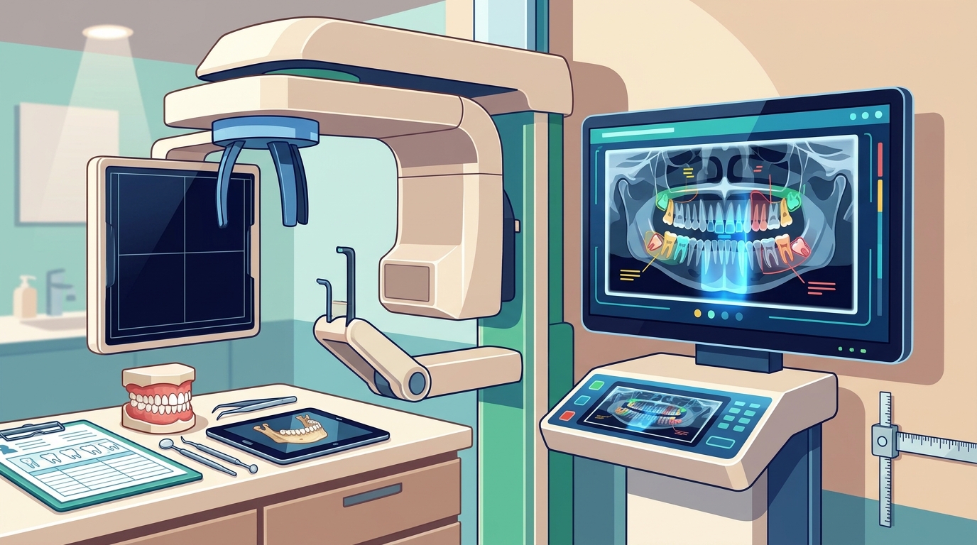
Digital X-rays also allow for the use of software tools that can highlight areas of interest. By zooming in on specific regions of the jaw, dentists can examine the trabecular pattern of the bone. Disruption in this pattern can be an early sign of aggressive pathology. This level of detail is crucial for differentiating between a benign cyst and a malignant tumor that has begun to erode the bone. The ability to review these images on a high-resolution monitor, rather than a physical film, ensures that no detail is overlooked during the screening process.
Radiation Dose Reduction
Patient safety is a paramount concern in any medical imaging procedure. Radiation dose reduction is a primary benefit of adopting digital X-ray technology over traditional film-based systems. Digital sensors are significantly more sensitive to X-rays, meaning they require a much lower exposure to produce a clear, diagnostic image.
Lower Exposure for Safer Screenings
Studies have consistently shown that digital radiography reduces radiation exposure by up to 80% compared to conventional film. This reduction is particularly important for patients who undergo regular dental check-ups and screenings. For individuals who may be at higher risk for oral cancer due to lifestyle factors or medical history, the ability to perform frequent screenings with minimal radiation exposure is a significant advantage. It allows for more vigilant monitoring without accumulating unnecessary radiation dose over time.
At our Herndon practice, we prioritize patient comfort and safety. By using digital X-rays, we ensure that every screening is as safe as possible. This is especially relevant for pediatric patients and those with specific health concerns. The lower dose not only reduces the theoretical risk of radiation-induced effects but also contributes to a more positive patient experience, as the procedure is often faster and more comfortable.
Environmental and Operational Benefits
While the primary benefit is patient safety, radiation dose reduction also has environmental and operational implications. Digital systems eliminate the need for chemical processing of film, reducing the environmental footprint of the practice. Furthermore, the immediate availability of digital images means that patients do not need to return for a second exposure if the initial image is not optimal. This efficiency ensures that the diagnostic process is streamlined, reducing the total number of exposures a patient might receive over the course of their dental care.
Biopsy Confirmation Role
While digital X-rays are powerful screening tools, they are not definitive diagnostic instruments for oral cancer. A definitive diagnosis requires a biopsy confirmation, where a sample of tissue is removed and examined under a microscope. However, digital X-rays play a crucial role in the biopsy process by guiding the clinician to the precise location of the lesion.
Guiding Precise Tissue Sampling
When a suspicious lesion is identified during a visual exam, the digital X-ray helps determine the depth and extent of the abnormality. This information is critical for planning the biopsy. If the lesion appears to involve the bone, the biopsy may need to be deeper or more extensive than a simple superficial swab. By providing a clear map of the underlying anatomy, digital X-rays ensure that the tissue sample taken is representative of the entire lesion, increasing the likelihood of an accurate diagnosis.
At Tooth Doc Family Dentistry, we believe in a collaborative approach to care. If a biopsy is indicated, we use the digital imaging data to communicate effectively with oral surgeons or oncologists. The ability to share high-quality digital images instantly with specialists ensures that they have the best possible information before performing the procedure. This coordination is essential for minimizing the number of biopsies needed and ensuring that the first sample is diagnostic.
Monitoring Post-Biopsy Healing
After a biopsy, digital X-rays are also used to monitor the healing process. They allow us to track the site of the biopsy over time, ensuring that the area is healing correctly and that there are no signs of residual disease. This ongoing monitoring is a key part of the comprehensive care we provide. By using digital X-rays for follow-up, we can detect any recurrence early, allowing for prompt intervention if necessary. This continuous loop of screening, diagnosis, and monitoring is what makes digital imaging an indispensable tool in oral cancer management.
Comparison of Imaging Modalities
| Feature | Traditional Film X-Ray | Digital X-Ray |
|---|---|---|
| Radiation Dose | Higher | Lower (up to 80% reduction) |
| Image Manipulation | Limited | Enhanced (zoom, contrast, brightness) |
| Turnaround Time | Delayed (processing required) | Immediate |
| Sharing with Specialists | Physical transfer or scanning | Instant digital transmission |
| Bone Detail Resolution | Standard | High (dynamic range) |
Key Takeaways
- Digital X-rays significantly enhance the detection of bone abnormalities associated with oral cancer.
- The use of digital sensors reduces radiation exposure by up to 80% compared to traditional film.
- Digital imaging provides the anatomical roadmap necessary for precise biopsy planning.
- Immediate image availability allows for faster diagnosis and treatment initiation.
- High-resolution digital images facilitate better communication between dentists and specialists.
- Regular screenings with digital X-rays are safer and more effective for long-term monitoring.
- At Tooth Doc Family Dentistry, we integrate these technologies into every routine exam.
- Early detection through advanced imaging is the most critical factor in oral cancer survival.
Frequently Asked Questions
Are digital X-rays safe for regular oral cancer screenings?
Yes, digital X-rays are considered very safe. They use a significantly lower dose of radiation than traditional film X-rays, making them ideal for routine screenings. The benefits of early detection far outweigh the minimal radiation risk associated with digital imaging.
Can a digital X-ray diagnose oral cancer on its own?
No, a digital X-ray cannot provide a definitive diagnosis of oral cancer. It is a screening tool that helps identify suspicious areas. A definitive diagnosis requires a biopsy, where tissue is examined under a microscope. However, the X-ray guides the biopsy process.
How often should I get a digital X-ray for oral cancer screening?
For most patients, a digital X-ray is recommended every 6 to 12 months as part of a routine dental exam. Patients with higher risk factors, such as a history of tobacco use or previous oral cancer, may need more frequent screenings. Your dentist will recommend a schedule based on your individual risk profile.
What is the difference between a panoramic X-ray and a bitewing X-ray?
A panoramic X-ray provides a broad view of the entire mouth, including the jawbones and sinuses. It is useful for detecting large abnormalities. A bitewing X-ray focuses on the upper and lower teeth, providing a detailed view of the crowns and the bone between them. Both can be used in oral cancer screening, depending on the area of concern.
Does digital X-ray technology work for children?
Yes, digital X-rays are safe and effective for children. The lower radiation dose is particularly beneficial for pediatric patients. At Tooth Doc Family Dentistry, we use gentle techniques and digital imaging to ensure that children's screenings are comfortable and safe.
Can I see the digital X-ray images during my appointment?
Yes, one of the major benefits of digital X-rays is that they can be displayed on a monitor in the exam room. This allows you to see exactly what your dentist is seeing, making it easier to understand your diagnosis and treatment options. We encourage patients to ask to see their images during their visit.
How does digital imaging help with biopsy confirmation?
Digital imaging helps by showing the depth and extent of a suspicious lesion. This information allows the dentist or surgeon to plan the biopsy more accurately, ensuring that the tissue sample taken is representative of the entire abnormality. This increases the likelihood of a correct diagnosis on the first attempt.
Is there any pain involved in getting a digital X-ray?
No, getting a digital X-ray is a painless procedure. It is similar to getting a traditional dental X-ray. The process is quick, and you will not feel any sensation from the X-ray itself. The only discomfort might be from holding your head still for a few seconds.
Conclusion
Digital X-rays have revolutionized oral cancer screening by providing a safer, more accurate, and more efficient diagnostic tool. The ability to detect bone abnormalities, reduce radiation exposure, and guide biopsy confirmation makes digital imaging an essential component of modern dental care. At Tooth Doc Family Dentistry, we are committed to using these advanced technologies to ensure that our patients receive the highest standard of care. If you are due for a dental exam or have concerns about your oral health, we encourage you to schedule an appointment with us. Our team is dedicated to helping you maintain a healthy, cancer-free smile.